Many of us have sat in a dental chair while the hygienist drapes that heavy lead apron over our chest for X-rays. Dental X-rays are powerful diagnostic tools that help spot potential problems before they become visible or painful.
While they might seem routine, your age, oral health history, and risk factors all play a role in determining how often you need these important scans.
Types of Dental X-Rays: Understanding Your Diagnostic Options
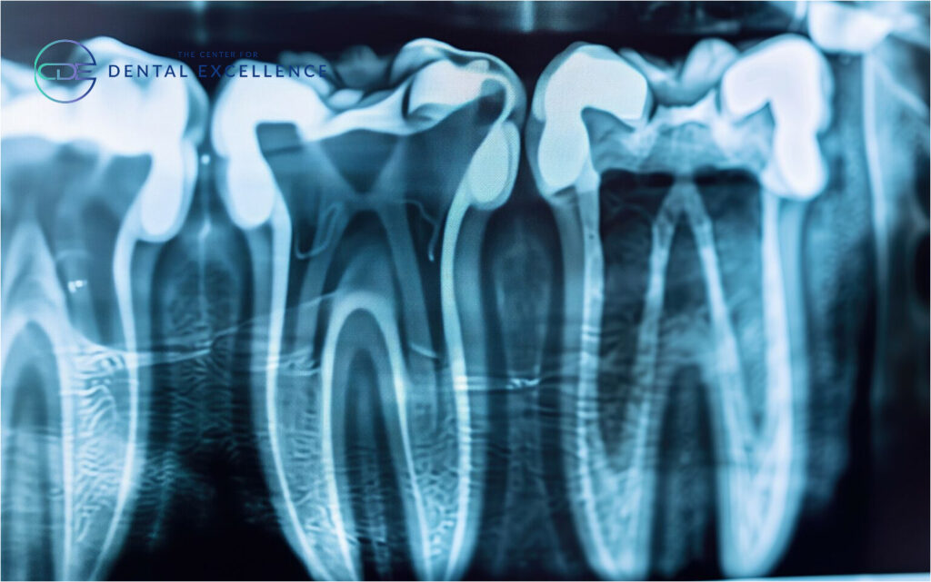
Bitewing X-rays are the most common type, capturing detailed images of your upper and lower teeth in the back of your mouth simultaneously. These dental X-rays detect cavities between teeth, evaluate bone density, and assess fillings. They’re typically taken yearly during check-ups and provide crucial information about decay that’s invisible to the naked eye.
Periapical X-rays capture an entire tooth from the crown to the root tip, including the surrounding bone structure. These detailed images help dentists diagnose root problems, evaluate deep tooth decay, identify bone loss from gum disease, and assess injuries. They’re essential before dental procedures like root canals or extractions.
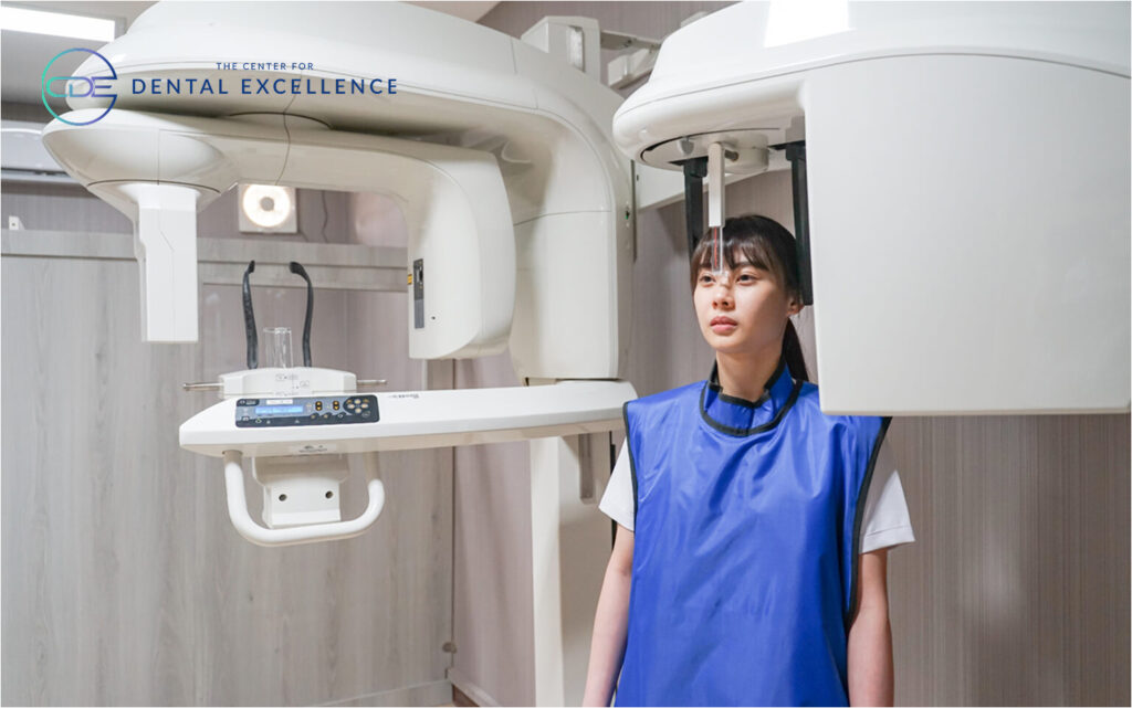
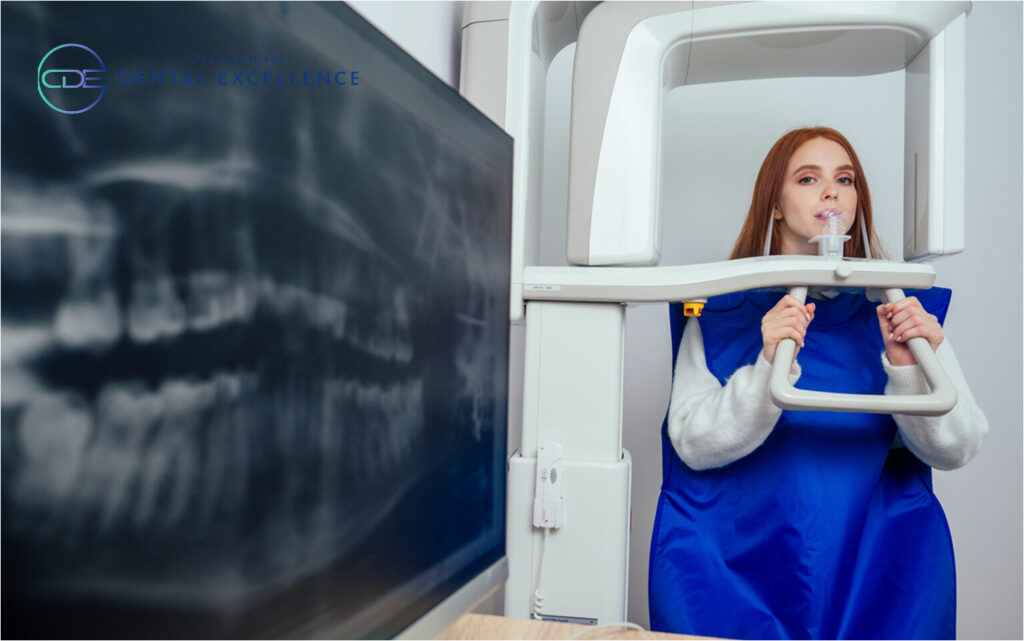
Panoramic X-rays provide a comprehensive view of your entire mouth in a single image, including teeth, jaws, sinuses, and surrounding structures. Unlike other X-rays, the machine rotates around your head while you stand still. Dentists use these to evaluate wisdom teeth, plan implant placement, detect cysts or tumors, and assess overall dental development in children.
Usage Guidelines:
- Bitewings: Annual check-ups, cavity detection
- Periapical: Root problems, pre-treatment planning
- Panoramic: Every 3-5 years, orthodontic planning, wisdom teeth evaluation
Each type serves distinct diagnostic purposes, and your dentist will recommend specific X-rays based on your dental health needs, age, and risk factors.
Standard X-ray Frequency Guidelines: What to Expect
When you first visit a new dentist, they’ll typically recommend a comprehensive baseline examination, including a full mouth series or panoramic X-ray, along with bitewing X-rays. This initial set creates a complete picture of your oral health and serves as a reference point for future visits.
For adult maintenance, healthy patients generally need bitewing X-rays every 12-18 months, with a full mouth series recommended every 3-5 years. However, this schedule may be adjusted based on your oral health history and current conditions.
Children require special consideration in X-ray scheduling. Most dentists begin taking X-rays when the child’s baby teeth start touching, typically around age 4-6. During cavity-prone years and periods of rapid development, more frequent imaging helps monitor tooth emergence and identify potential problems early. Panoramic X-rays become particularly important during orthodontic planning phases.
Certain risk factors may necessitate more frequent X-rays. These include a history of extensive decay, gum disease, smoking, dry mouth conditions, diabetes, or numerous existing dental work. Your dentist will assess these factors to create a personalized imaging schedule that balances diagnostic needs with radiation exposure considerations. The American Dental Association’s guidelines serve as a framework, but your specific schedule will depend on your individual oral health needs.
Factors That Determine Your X-ray Schedule
Understanding the variables that influence X-ray timing helps explain why your dentist may recommend different imaging schedules for different patients.
Age-Related Factors
Age determines X-ray frequency, with children needing more frequent imaging to monitor tooth development and orthodontic needs. Adults with stable oral health require less frequent X-rays, while seniors often need increased monitoring for root decay and bone density changes.
Oral Health Background
Past dental issues like frequent cavities, periodontal disease, or multiple restorations require closer monitoring through X-rays. Your history helps dentists establish appropriate imaging intervals to protect existing work and prevent new problems.
Current Dental Status
Active dental problems like pain, visible decay, or changes in bite often necessitate immediate X-rays. Your dentist may require additional images to diagnose specific issues or monitor treatment progress.
Medical Conditions
Systemic conditions affect oral health significantly. Diabetes, autoimmune disorders, and medications causing dry mouth can accelerate dental problems. Cancer treatment history, especially radiation to the head or neck, requires careful monitoring through regular X-rays.
Genetic Considerations
A family dental history of early tooth loss, aggressive periodontal disease, or frequent decay may indicate a genetic predisposition to dental problems. This information helps determine appropriate X-ray intervals for preventive dental care.
Safety and Modern Technology in Dental X-rays
Modern dental imaging technology has revolutionized patient safety while enhancing diagnostic capabilities. Digital X-rays reduce radiation exposure by up to 80% compared to traditional film, providing instant results and superior image quality for better diagnosis.
The radiation exposure from dental X-rays is remarkably low. A full set exposes patients to less radiation than a cross-country flight, with digital bitewing X-rays delivering about 0.005 millisieverts – equivalent to one day of natural background radiation. Modern equipment uses focused beams to minimize scatter radiation.
Dental offices employ comprehensive safety protocols, including lead aprons, thyroid collars, and beam-limiting devices. Digital sensors require shorter X-ray exposure times, while regular equipment calibration and staff training ensure optimal safety standards are maintained.
Pregnancy requires special consideration in dental imaging. While X-rays are generally safe during pregnancy, dentists typically postpone routine imaging until after delivery. For emergency situations requiring X-rays, additional precautions like double lead apron protection are implemented. The American Dental Association confirms that necessary dental X-rays pose minimal risk when proper safety measures are followed.

Signs You May Need Additional X-rays
When tooth pain or sensitivity strikes, it often signals problems hiding beneath the surface. Whether you’re experiencing sharp pain while eating ice cream or a persistent ache when chewing, X-rays help your dentist pinpoint the exact cause and extent of the issue.
Visible changes in your teeth or gums shouldn’t be ignored. Dark spots, chips, or obvious cavities are like warning flags, but the visible damage may only be the tip of the iceberg. X-rays reveal how deep the problem extends and guide your dentist in planning the most effective dental treatment.
Your gums tell an important story about your oral health. When they bleed, recede, or become swollen, it could indicate serious problems developing below the surface. X-rays are crucial for evaluating bone health and detecting early signs of periodontal disease before it progresses too far.
Dental emergencies demand immediate attention and almost always require X-rays. Whether you’ve taken a hit to the mouth during sports or woken up with severe swelling, these images provide critical information about potential fractures, infections, or root damage that might not be visible to the naked eye. Quick imaging in emergency situations helps your dentist make informed decisions about urgent treatment needs.
Make Informed Decisions About Your Dental X-rays
Regular dental X-rays are a crucial part of maintaining optimal oral health, helping detect problems before they become serious complications. Your dentist will create a personalized imaging schedule based on your unique risk factors, oral health history, and current dental status.
Remember that modern digital X-rays combine safety with superior diagnostic capabilities, making them an invaluable tool in preserving your bright, healthy smile.